Pulmonary fibrosis is a debilitating and deadly disease that requires accurate diagnosis for directing appropriate treatment, often requiring an invasive surgical biopsy.

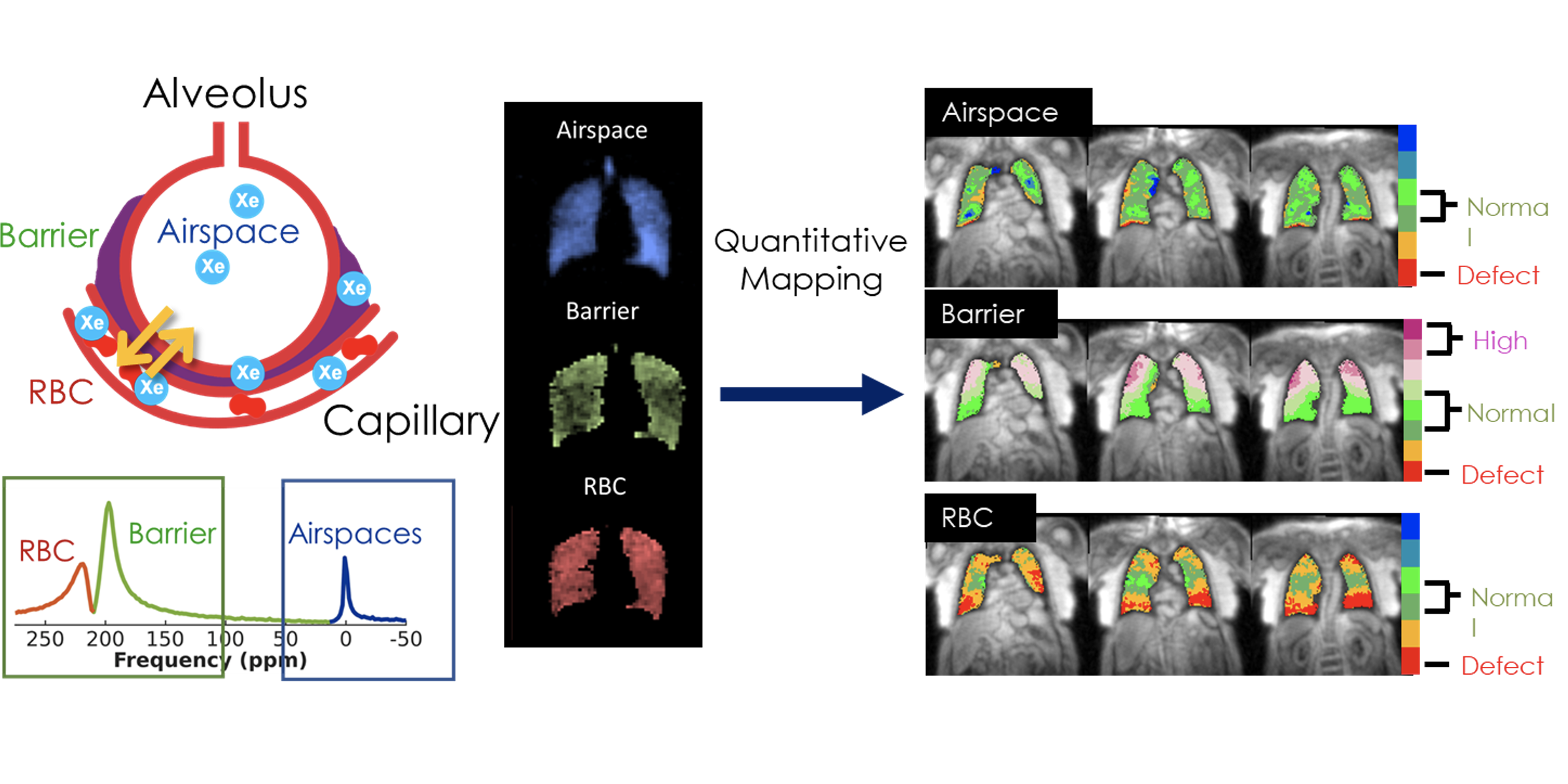
Dr. Joseph Mammarappallil, in collaboration with Dr. Bastiaan Driehuys and a team of researchers, have applied a novel approach to characterizing pulmonary fibrosis using an inhaled gas (hyperpolarized 129 Xe) and MRI.

This imaging technique allows visualization of diseased portions of the lung not detectable by CT or other imaging methods, potentially allowing for earlier diagnosis.
Furthermore, this imaging technique may help differentiate among the various subtypes of fibrotic interstitial lung disease, which could prevent the need for an invasive and risky surgical procedure. In addition to earlier and more accurate diagnosis, MRI with hyperpolarized gas also promises to be an important study for monitoring disease progression and treatment efficacy. This imaging technique could theoretically identify patients who are responsive to medications while also transitioning patients to more effective therapies to more effectively prevent disease progression. Dr. Mammarappallil and his team are exploring additional applications of this advanced imaging technology which may be beneficial for other types of chronic lung disease.