RESEARCH
CURRENT CLINICAL TRIALS
Randomized trial of CT fluoroscopy-guided targeted autologous blood and fibrin glue patching for treatment of cerebrospinal fluid leaks in spontaneous intracranial hypotension (SIH).
Duke investigators are conducting the first prospective randomized clinical trial assessing the efficacy of CT fluoroscopy-guided targeted blood and fibrin glue patching of confirmed CSF leaks in patients with spontaneous intracranial hypotension. An outcome of equivalent or inferior efficacy would result in a paradigm shift for SIH treatment (i.e. targeted patching would no longer be considered optimal therapy). Demonstrated superior efficacy would validate this procedure leading to a substantial increase in the performance of targeted patching and would provide the basis for future research comparing its efficacy with other epidural blood patch techniques. Patients interested in enrolling may contact our team for further information.
CONTACT:
Timothy J. Amrhein, M.D. (Principal Investigator)
919-684-7770
Assistant Professor of Radiology
Division of Neuroradiology
Department of Radiology
Duke University Medical Center
EDUCATION
UPCOMING COURSES:

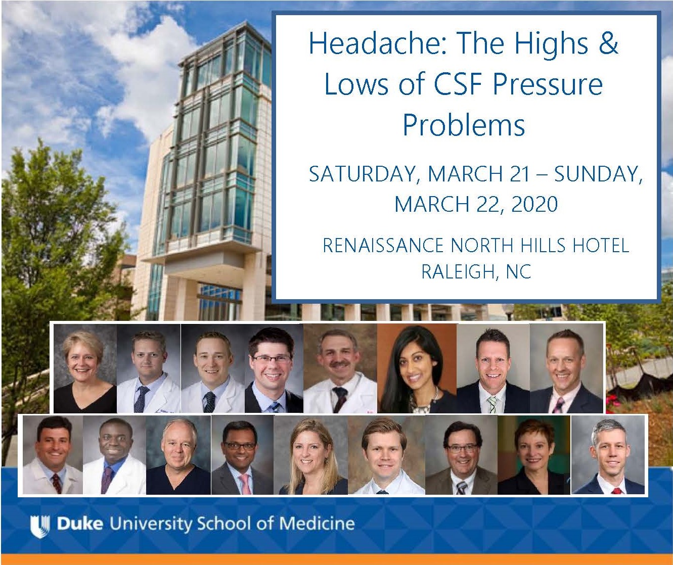
This 1½-day multidisciplinary course on Intracranial Hypotension secondary to spinal cerebrospinal fluid (CSF) leak is intended to inform local neurologists, headache specialists, and other health care professionals including neurosurgeons, anesthesiologists, neuroradiologists, and pain management physicians about the varied presentations and the current state of diagnosis and treatment for this disorder. Intracranial Hypotension can present as migraine, chronic migraine, new daily persistent headache, and Chiari I malformations and can be iatrogenic in origin as can be seen with lumbar punctures, epidural catheter placement and epidural injections. The approach to the diagnosis with CT guided lumbar puncture, myelogram, and minimally invasive techniques including fibrin glue and bloodpatching as well as indications for surgical intervention will be discussed. Invited speakers include neuroradiology, neurology-ophthalmology, neurosurgery, and orthopedic surgery. If you have cases you would like to have discussed during a last session, send cases for consideration ahead of time to Dr. Gray Leithe at linda.leithe@duke.edu.
HOW YOU CAN HELP
Although this condition is being recognized more frequently, much more work needs to be done. With your help, we can continue our groundbreaking efforts to:
UNDERSTAND THE DISEASE BETTER
There still remain many unanswered questions about why this condition develops, how the symptoms develop and change with time, and how the process of recovery occurs after treatment. Research requires teams of collaborators, scientists, statisticians, and research assistants, and the funding to support them.
FIND THE MOST EFFECTIVE TREATMENT
There are very few studies in the medical literature that look at the different treatment options for this condition and which ones work best. We need this information desperately. We are currently conducting high-quality trials of treatment outcomes, but these trials are expensive, labor-intensive, and need financial support to be successful.
TRAIN MORE PHYSICIANS TO TREAT THE CONDITION
As more and more patients are diagnosed , more centers are needed to provide high-quality treatment. Although we currently teach physicians-in-training and host visiting practicing physicians, greater funding could establish dedicated training programs such as a fellowship in this area.
EDUCATE PHYSICIANS AROUND THE COUNTRY AND WORLD
We need to educate other healthcare providers on how to recognize this condition. One of the best ways to do this is provide education and share our experience by traveling to meetings, conferences, and hospitals as well as through educational information published in highly respected medical journals. We have already seen the benefits of programs such as these and are often asked to consult with physicians from inside the US and around the world. We need your help to continue this outreach.
DISCOVER
Every day we make new discoveries about this condition, and often these discoveries are unexpected. We treat patients differently today than we did 10, 5 or even 2 years ago as a result of these discoveries. We need help to follow these leads when they arise, as they often result in giant leaps forward in our knowledge.
DONATIONS
Sally Schatz, Director of Development
Office: 919-385-0034
Fax: 919-385-3103
Email: sally.schatz@duke.edu
Location: Duke Health Development and Alumni Affairs
300 W. Morgan St., #1200, Durham, NC 27701
Durham, NC 27701
PUBLICATIONS
- Kranz PG, Gray L, Taylor JN. CT-Guided Epidural Blood Patching of Directly Observed or Potential Leak Sites for the Targeted Treatment of Spontaneous Intracranial Hypotension. AJNR Am J Neuroradiology. 2011 May;32(5):832-8. doi:10.3174/ajnr.A2384. Epub 2011 Feb 24. PMID: 21349964
- Kranz PG, Viola RJ, Gray L. Resolution of syringohydromyelia with targeted CT-guided epidural blood patching. J Neurosurg. 2011 Sep;115(3):641-4. doi:10.3171/2011.3.JNS102164. Epub 2011 Apr 29. PMID: 21529136
- Kranz PG, Stinnett SS, Huang KT, Gray L. Spinal meningeal diverticula in spontaneous intracranial hypotension: analysis of prevalence and myelographic appearance. AJNR Am J Neuroradiol. 2013 Jun-Jul;34(6):1284-9. doi:10.3174/ajnr.A3359. Epub 2012 Dec 6. PMID: 23221945.
- Kranz PG and Provenzale JM. Imaging “worst headache of my life” Part 1: Conditions in which the initial CT is often positive. Applied Radiology, 2013 Jul; 42(7):19-25.
- Kranz PG and Provenzale JM. Imaging “worst headache of my life” Part 2: Conditions where initial CT is often normal, but other imaging may be diagnostic. Applied Radiology. 2013 Aug: 12-16.
- Kranz PG, Amrhein TJ, Gray L. Rebound intracranial hypertension: a complication of epidural blood patching for intracranial hypotension. AJNR Am J Neuroradiol. 2014 Jun;35(6):1237-40. doi:10.3174/ajnr.A3841. Epub 2014 Jan 9. Review. PMID: 24407273.
- Mihlon F, Kranz PG, Gafton AR, Gray L. Computed tomography-guided epidural patching of postoperative cerebrospinal fluid leaks. J Neurosurg Spine. 2014 Nov;21(5):805-10. doi:10.3171/2014.7.SPINE13965 Epub 2014 Aug 15. PMID: 25127431.
- Howard BA, Gray L, Isaacs RE, Borges-Neto S. Definitive diagnosis of cerebrospinal fluid leak into the pleural space using 111ln-DTPA cisternography. Clin Nucl Med 2015 40(3):220-3.
- Kranz PG, Tanpitukpongse TP, Choudhury KR, Amrhein TJ, Gray L. How common is normal cerebrospinal fluid pressure in spontaneous intracranial hypotension? Cephalalgia. 2015 Dec 17. pii: 0333102415623071 [Epub ahead of print]. PMID:668257540(3):220-3.
- Kranz PG, Luetmer PH, Diehn FE, Amrhein TJ, Tanpitukpongse TP, Gray L. Myelographic Techniques for the Detection of Spinal CSF Leaks in Spontaneous Intracranial Hypotension. AJR Am J Roentgenol. 2016 Jan; 206(1):8-19. doi:10.2214/AJR.15.14884. Review. PMID:26700332
- Capizzano AA, Lai L, Kim J, Rizzo M, Gray L, Smoot K, Moritani T. Atypical presentations of intracranial hypotension: Comparison with classic spontaneous intracranial hypotension. AJNR Am J Neuroradiol. 2016 (March 3), PMID 26939631.
- Kranz PG, Amrhein TJ, Schievink WI, Karikari IO, Gray L. The “Hyperdense Paraspinal Vein” Sign: A Marker of CSF-Venous Fistula. AJNR Am J Neuroradiol. 2016 Jul; 37(7):1379-81. doi:10.3174/ajnr.A4682. PMID:26869470
- Kranz PG, Tanpitukpongse TP, Choudhury KR, Amrhein TJ, Gray L. Imaging Signs in Spontaneous Intracranial Hypotension: Prevalence and Relationship to CSF Pressure. AJNR Am J Neuroradiol. 2016 Jul; 37(7):1374-8. doi:10.3174/ajnr.A4689. PMID: 26869465
- Amrhein TJ, Befera NT, Gray L, Kranz PG. CT Fluoroscopy-Guided Blood Patching of Ventral CSF Leaks by Direct Needle Placement in the Ventral Epidural Space Using a Transforaminal Approach. AJNR Am J Neuroradiol. 2016 Jul 7. [Epub ahead of print] PMID: 27390315
- Kranz PG, Amrhein TJ, Choudhury KR, Tanpitukpongse TP, Gray L. Time-Dependent Changes in Dural Enhancement Associated With Spontaneous Intracranial Hypotension. AJR Am J Roentgenol. Epub 2016 Aug 24:1-5. AJR Am J Roentgenol. 2016 Dec; 207(6):1283-1287. PMID: 27557149
- Kranz PG, Malinzak MD, Amrhein TJ, Gray L. Update on the Diagnosis and Treatment of Spontaneous Intracranial Hypotension. Curr Pain Headache Rep. 2017 Aug;21(8):37. doi: 10.1007/s11916-017-0639-3. Review. PMID: 28755201
- Kranz PG, Amrhein TJ, Gray L. CSF Venous Fistulas in Spontaneous Intracranial Hypotension: Imaging Characteristics on Dynamic and CT Myelography. AJR Am J Roentgenol. 2017 Oct 12:1-7. doi: 10.2214/AJR.17.18351. [Epub ahead of print] PMID: 29023155
- Kranz PG, Gray L, Amrhein TJ. Spontaneous Intracranial Hypotension: 10 Myths and Misperceptions. Headache. May 2018.
- Kranz PG, Malinzak MD, Amrhein TJ. Approach to Imaging in Patients with Spontaneous Intracranial Hemorrhage. Neuroimaging Clin N Am. 2018 Aug;28(3):353-374. doi: 10.1016/j.nic.2018.03.003. Epub 2018 Jun 8. Review.
- Kranz PG, Gray L, Amrhein TJ. Decubitus CT Myelography for Detecting Subtle CSF Leaks in Spontaneous Intracranial Hypotension. AJNR Am J Neuroradiol. 2019 Feb 28. doi: 10.3174/ajnr.A5995. [Epub ahead of print] PMID: 30819772
- Amrhein TJ, Kranz PG. Spontaneous Intracranial Hypotension: Imaging in Diagnosis and Treatment. Radiol Clin North Am. 2019 Mar;57(2):439-451. doi: 10.1016/j.rcl.2018.10.004. Epub 2018 Dec 7. Review. PMID: 30709479
- Kranz PG, Amrhein TJ. Imaging Approach to Myelopathy: Acute, Subacute, and Chronic. Radiol Clin North Am. 2019 Mar;57(2):257-279. doi: 10.1016/j.rcl.2018.09.006. Epub 2018 Dec 5. Review. PMID: 30709470
- Wang TY, Karikari IO, Amrhein TJ, Gray L, Kranz PG. Clinical Outcomes Following Surgical Ligation of Cerebrospinal Fluid-Venous Fistula in Patients With Spontaneous Intracranial Hypotension: A Prospective Case Series. Oper Neurosurg (Hagerstown). 2019 May 28 [Epub ahead of print].
