THE PROCEDURE
The lesion to be treated is first localized by either CT or ultrasound. At times, both CT and ultrasound are used. A corresponding mark is made with a felt tip pen on the skin which is then cleansed with a cold soap (Betadine) and a large plastic drape is placed over the cleansed area to maintain a sterile field.

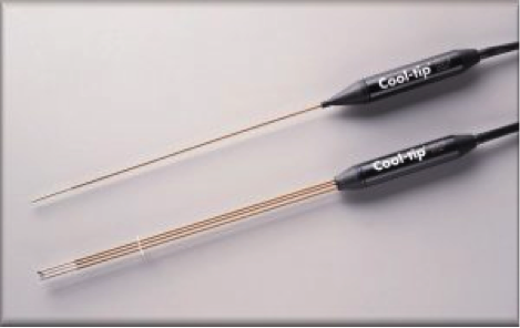
xample of probes inserted into a tumor for deposition of RF energy. The top devise is a single probe which is used for small lesions. The bottom device is a triple or “cluster” probe which is used for larger lesions. These probes have “Cool-tip” stenciled on the handle because cold water is circulated inside the probes to increase the amount of tissue destroyed.
For pain control, both local and intravenous medications will be administered and is addressed in detail below. One or more tiny incisions are then made to the skin, each measuring less than 1/4 inch in length. The Radiofrequency, Microwave, or Cryoablation probes are then advanced into the lesion using CT, ultrasound, or both to guide placement of the probe. Once in place, the probe is hooked up to a special machine which controls and monitors the production of heat in the case of RF and MW and the production of an ice ball in the case of Cryoablation. This process requires several minutes (up to 30 minutes per ablative session), depending upon the size of the lesion being treated.
Larger lesions can take longer or require multiple treatment sessions. Since it is our goal to destroy both the tumor and a cuff of normal tissue around the tumor, we often treat each lesion more than once within a single session.
When the procedure is complete and the probe(s) removed, a Band-Air or gauze dressing will be placed over the incision(s). The patient will be moved to the Radiology recovery room for 2 – 4 hours of observation and released at the point when all of the following conditions are met:
- Vital signs stable: lying down, sitting up, and standing
- Adequate pain control
- Able to tolerate liquids and solids by mouth
- Able to empty the bladder
- No signs of complications
RISKS
Anytime a needle is placed under the skin there is always the risk of bleeding and/or infection. Prior to the procedure, blood work will be done to check for a bleeding tendency. Bleeding complications are further minimized by “coagulating” the tract with Radiofrequency or Microwave energy upon withdrawal of the probe.
Infectious complications are minimized by administering antibiotics both intravenously during the procedure and by mouth for several days after the procedure.
Other less common complications vary depending upon the site of treatment. Diaphragmatic injury may occur with kidney, liver, or lung ablation and often manifests as right shoulder pain.
The lung may collapse during the treatment of lung tumors, or lesions within the liver that are adjacent to the diaphragm. Lung collapse may require placement of a small tube between the lung and chest wall to re-inflate the lung.
A leak of bile or urine can occur when treating kidney or liver lesions, respectively.
On rare occasions, significant damage to the liver or kidney may occur as a results of the ablation. If the severity of organ damage is significant, the injury may be life-threatening.
Injury to other structures such as blood vessels or the bowel is unlikely when CT or US are used to guide probe placement. Experience has shown that all of these complications are uncommon, occurring in approximately 5% or less of the patients.