Genetics plays an outsized role in determining who develops Alzheimer’s disease but many people with genes linked to the disease don’t develop symptoms.
To better understand why some people are more vulnerable while others are resilient, researchers from Duke University and the University of Tennessee Health Science Center (UTHSC) used ultra-high-resolution magnetic resonance (MR) microscopy to map volume changes throughout the brain in a carefully crafted collection of genetically engineered mouse models. The results provide new insight into the evolution of the disease and make it easier for scientists to test potential Alzheimer’s treatments in preclinical trials.
The researchers — including Duke’s G. Allan Johnson, Ph.D., Charles E. Putman University Professor of Radiology, Physics and Biomedical Engineering; Robert W. Williams, Governor's Chair of Computational Genomics at UTHSC; and David G. Ashbrook, now at the University of Edinburgh — published their findings in Nature Neuroscience in February 2026.
“MRI has revolutionized our ability to follow the impact of neurodegenerative disease on brain structure,” Johnson said. “But clinical studies are enormously complex and costly. The randomness of human genetics forces clinical studies to include up to 50,000 patients to achieve statistical robustness.
“Preclinical studies with replicable genetic models allow us to complete statistically significant studies with hundreds of animals. The problem has been achieving adequate resolution for the much smaller mouse brain. The Duke mouse connectome scanner has removed this barrier.”
The researchers studied a unique mouse population that featured Alzheimer’s-linked genes in a widely used mouse model (5×FAD). The mice had genetic diversity similar to humans, allowing the team to test how different genetic backgrounds can affect brain changes caused by Alzheimer’s-related genes. By comparing mice with the mutations to those without, the researchers created a highly detailed map of volume changes across the brain, showing the impact of those mutations on the brain across multiple backgrounds.
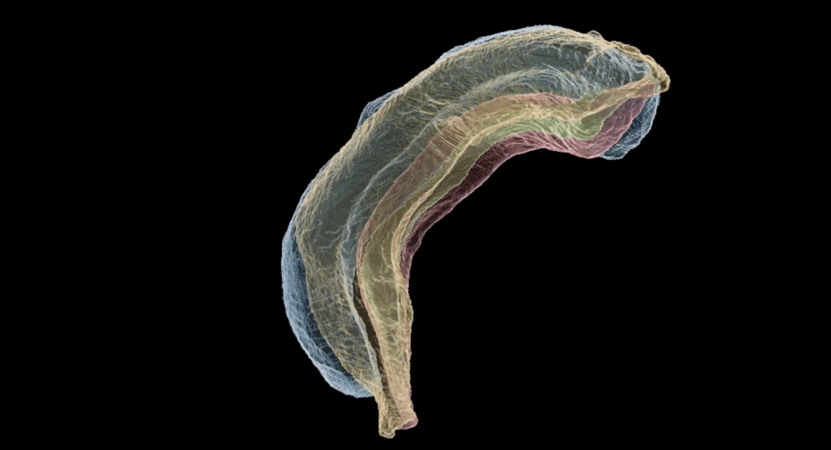
The MR microscope they used — which was developed at Duke — featured a spatial resolution about 500,000 times finer than a typical clinical MRI. They used diffusion tensor imaging (DTI) on postmortem brain specimens to follow changes in 230 regional volumes covering the entire brain. DTI measures how water moves through brain tissue, making it highly sensitive to the brain’s microscopic structure and connection pathways. Using high performance computing, they create a detailed map of volume changes across the entire brain — where the brain swelled and shrank — showing the impact of Alzheimer’s genes and the background on that map.
The researchers found that the background genome impacts who develops Alzheimer’s. Imaging showed changes were large for some genetic backgrounds and statistically insignificant for others. Over half of the regions changed, though total brain volume was unchanged. They also found that female mice had higher variability in volumes. Importantly, these neuroanatomical changes correlated with changes in memory and learning behaviors traits relevant to Alzheimer’s disease symptoms.
“The Duke/UTHSC collaboration has been ongoing for nearly 20 years,” Johnson said. “We have established a unique partnership in genetics, neuroscience, engineering, and computer science that has positioned us for substantive breakthroughs in following neurodegenerative disease.”