
Faculty Network Member of the Duke Institute for Brain Sciences
Overview
The Duke Positron Imaging Research Lab (PIRL) provides PET, PET/CT, and CT imaging for human and large animal research. It is a School of Medicine service center operated in the Department of Radiology.
PET (positron emission tomography) allows a wide array of biological mechanisms to be studied through the administration of a small (~nanogram) quantity of a radioactive tracer and a subsequent scan to determine where in the body the tracer went (quantitatively) and, in some cases, its time course. PET imaging has been used heavily in oncologic, cardiac, neurologic, and other applications.
The Lab’s imaging services are appropriate for multicenter trials and investigator-initiated human and animal research. Those interested in research involving PET should contact us early in the process to discuss the research needs and available methods, determine feasibility, and develop proposals.
Some PET research allows (and requires) substantial image analysis. Our lab can provide that service, or assist investigators equipped to do the analysis themselves.
Some research is better suited for the clinical PET/CT facility (for a variety of reasons). The PIRL staff, Nuclear Medicine personnel and the Radiology practice manager will work with research teams to determine where best to do their work. Even for research performed on the clinical PET/CT scanners, the PIRL team provides all necessary extra support not typically provided for clinical scanning, allowing for successful outcomes even for complex protocols.
WHAT ARE WE SEEING?
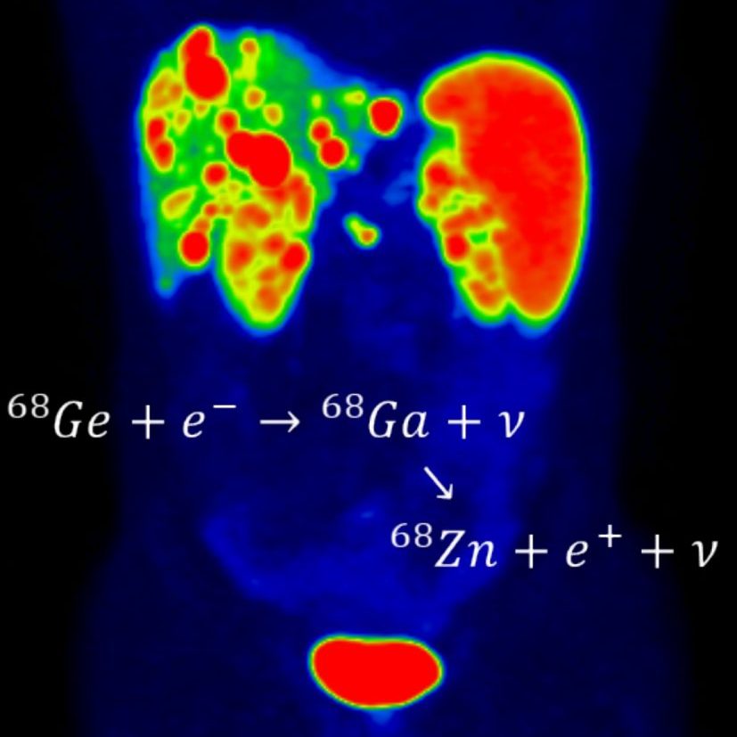
Each PET scan requires the administration (injection, inhalation, or injestion) of a specific 'radiotracer'. Radiotracers are designed to reveal specific biological properties of the body ranging from 'plumbing' questions like blood flow, to glucose utilization, and more specific cellular processes. Sometime after the administration (in some cases immediately, in other cases over an hour) the scan proceeds, revealing the distribution of the radioactive material in the body and therefore the underlying processes. Several radiotracers are available for routinely for use in humans, and the list is growing.
Radiotracers
Commercially and internally available (for human use)
FDG –Glucose Metabolism
Dotatate –Neuroendocrine Tumors
Axumin –Prostate Cancer
NaF –Bone Lesions
13NH3 –Cardiac Blood Flow
Florbetapir – beta amyloid
Florbetabin –beta amyloid
MK-6240 - tau tangles
68Ga PSMA – Prostate Cancer
The list of commercially available tracers changes frequently, and other tracers may be produced in-house for animal research. Please contact us with any questions on requests.
Specific Services
Subject (patient, volunteer, and animal) preparation and administration of radiotracer.
PET, PET/CT, and CT scan.
Image reconstruction and processing.
Anonymization and data uploading/delivery.
Special one-time or periodic PET/CT scanner calibrations, QC, or other qualification requirements unique to specific research projects.
Preparation of estimates and invoices for clients.
Assistance in audits.
Assistance in procurement of individualized radiopharmaceutical doses.
Assistance in scheduling scans and coordinating of the PET scans with any radiopharmacy needs.
EXAMPLE: CARDIAC PET
After the injection of the radiotracer (in this case [N-13] ammonia) its transit through the body is seen here in one cross-sectional slice over time. After injection in the arm, the material is next seen in the right ventricle, then the left ventricle (after a brief visit in the lungs), and on out to the body through the arteries. Some of this returns to the heart muscle itself. The process is displayed several times (in changing colors) then the beating heart (images from after the radiotracer has returned to the heart muscle is depicted - primarily the left ventricle but also (to the left) the right ventricle). This is one of many uses of PET.
EQUIPMENT AND SPACE
The lab is South Hospital, Yellow Zone, basement, near Yellow Elevator B, directly behind the HLD facility.
The primary equipment of the lab is a GE Discovery 690 PET/CT. This time-of-flight PET system with 16 cm axial field of view couple with a 64-slice VCT CT provides excellent images and quantitative measurements for a wide range of applications, and all common PET procedures. It has flexible scanning and post-processing capabilities allowing for novel applications.
In addition to the imaging room suite, there is ample space for patient/subject preparation, radioactive material storage and image processing. In addition to the scanner, there are image viewing and analysis workstations.
Contact Us
PERSONNEL
Director: Tim Turkington, PhD 919-684-7706 timothy.turkington@duke.edu
Medical Director: Terry Wong, MD, PhD, 919-684-7748 terence.wong@duke.edu
Thomas Hawk, Associate in Research 919-684-7712 thomas.hawk@duke.edu
Robin Davis, Associate in Research 919-684-7807 robin.davis@duke.edu
Priti Patel, NM Technologist 919-684-7714 priti.patel@duke.edu
For general communication, including scheduling scans, please send messages to lab shared email account pirl@duke.edu.